 NEW: Precision veterinary medicine
Guest Edited by Jose J. Ceron and
Carlos E. Fonseca-Alves
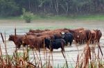 Climate change and animal health and welfare: informing an action agenda
Guest Edited by Colleen Duncan, Chris Oura, and Craig Stephen
 Veterinary public health and food security
Guest Edited by Mahdi Askari Badouei
Advan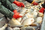 cements in veterinary medicine and aquaculture
Guest Edited by Hanan Hassan Abd-Elhafeez and Mohamed Shaalan
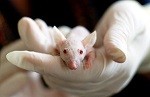
 Comparative medicine
Guest edited by Maria Grazia Cappai and Enrico Gugliandolo
 Mastitis and milk quality in dairy ruminants
Guest edited by Abdelfattah Selim and Fernando Nogueira de Souza
 Advances in canine health research
Guest edited by Selwyn Arlington Headley and Jelena Prpić
 Animal welfare and ethics
Guest edited by Izhar Hyder Qazi and Gemma Walmsley