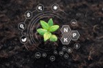
 Climate-smart soils to enhance sustainable crop yield
Guest Edited by Walter Mupangwa, Debadatta Sethi and Muhammad Shaaban
 Omics technologies and applications in horticultural crops
Guest Edited by Yunpeng Cao and Mohammad Shah Jahan
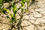 Crop breeding for drought stress tolerance
Guest Edited by Mostafa Abdelrahman, Ahmad M. Alqudah, Vijay Gahlaut and Daoquan Xiang
 Plant synthetic biology: advances in fundamental research and applications
Guest Edited by Jemaa Essemine, Saroj Kumar Sah and Qinlong Zhu