Complementary approaches to women's health
Guest Edited by Junyoung Jo, Fan Qu and Fangfang Wang
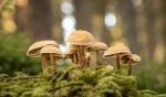 Advances in mycotherapy
Guest Edited by Wong Kah Hui and I-Chen Li
 Anxiety and stress-related disorders: the role of botanicals
Guest Edited by Ravid Doron, Yunna Kim and Wenda Xue
 Herb-drug interactions
Guest Edited by Awodayo O. Adepiti, Swapnil P. Borse, Márcio Rodrigues and Cheng-Peng Sun