- Case Report
- Open access
- Published:
A challenging case of carbapenem resistant Klebsiella pneumoniae-related pyogenic liver abscess with capsular polysaccharide hyperproduction: a case report
BMC Infectious Diseases volume 24, Article number: 433 (2024)
Abstract
Background
Carbapenem-resistant Klebsiella pneumoniae (CRKP) infections are a major public health problem, necessitating the administration of polymyxin E (colistin) as a last-line antibiotic. Meanwhile, the mortality rate associated with colistin-resistant K. pneumoniae infections is seriously increasing. On the other hand, importance of administration of carbapenems in promoting colistin resistance in K. pneumoniae is unknown.
Case presentation
We report a case of K. pneumoniae-related pyogenic liver abscess in which susceptible K. pneumoniae transformed into carbapenem- and colistin-resistant K. pneumoniae during treatment with imipenem. The case of pyogenic liver abscess was a 50-year-old man with diabetes and liver transplant who was admitted to Abu Ali Sina Hospital in Shiraz. The K. pneumoniae isolate responsible for community-acquired pyogenic liver abscess was isolated and identified. The K. pneumoniae isolate was sensitive to all tested antibiotics except ampicillin in the antimicrobial susceptibility test and was identified as a non-K1/K2 classical K. pneumoniae (cKp) strain. Multilocus sequence typing (MLST) identified the isolate as sequence type 54 (ST54). Based on the patient’s request, he was discharged to continue treatment at another center. After two months, he was readmitted due to fever and progressive constitutional symptoms. During treatment with imipenem, the strain acquired blaOXA−48 and showed resistance to carbapenems and was identified as a multidrug resistant (MDR) strain. The minimum inhibitory concentration (MIC) test for colistin was performed by broth microdilution method and the strain was sensitive to colistin (MIC < 2 µg/mL). Meanwhile, on blood agar, the colonies had a sticky consistency and adhered to the culture medium (sticky mucoviscous colonies). Quantitative real-time PCR and biofilm formation assay revealed that the CRKP strain increased capsule wzi gene expression and produced slime in response to imipenem. Finally, K. pneumoniae-related pyogenic liver abscess with resistance to a wide range of antibiotics, including the last-line antibiotics colistin and tigecycline, led to sepsis and death.
Conclusions
Based on this information, can we have a theoretical hypothesis that imipenem is a promoter of resistance to carbapenems and colistin in K. pneumoniae? This needs more attention.
Background
Pyogenic liver abscess (PLA) is a rare infectious disease usually caused by bacterial infection of the liver parenchyma in immunocompromised patients [1]. Klebsiella pneumoniae and Escherichia coli are the most common etiological agents of PLAs in immunocompromised patients [2, 3]. Liver transplantation and diabetes mellitus (DM) are two known risk factors for the development of PLA [1, 4]. K. pneumoniae readily colonizes the intestine and can cross the intestinal barrier to enter the liver via the portal vein system and cause PLA in healthy and immunocompromised individuals [1, 2]. Antibiotic resistance in K. pneumoniae arises from several mechanisms, including enzymes that directly inactivate antibiotics (e.g., β-lactamases or aminoglycoside-modifying enzymes), activation of efflux pumps that pump out the antibiotic, and mutations in the antibiotic target [5]. The prevalence of multidrug-resistant K. pneumoniae strains producing carbapenemase is increasing [6]. OXA-48 is one of the most common carbapenemases and is frequently detected in K. pneumoniae and E. coli strains [7]. Treatment of infection caused by carbapenem-resistant K. pneumoniae (CRKP) isolates is often limited to colistin as a last-line antibiotic [8]. However, the high mortality rate of CRKP infections due to limited treatment options indicate the urgent need for new strategies to treat K. pneumoniae infections [8].
Capsular polysaccharide is one of the most important virulence factors of K. pneumoniae [9]. Genes involved in capsule production are located on the cps operon (capsular polysaccharide synthesis) of the chromosome [9]. The expression of this gene cluster (from galF to ugd) is driven by three promoters located upstream of galF, wzi, and manC genes, respectively. The conserved galf, cpsACP, wzi, wza, wzb and wzc genes at the 5’ end of the cps gene cluster are involved in the transfer and processing of capsular polysaccharide at the bacterial surface [9]. The Rcs (regulator of capsule synthesis) system is a cell envelope stress response system and is present in many Enterobacterales species [10]. The Rcs system is activated by outer membrane damage, lipopolysaccharide (LPS) synthesis defects, and peptidoglycan disruption and regulates the expression of genes involved in capsule biosynthesis, motility, biofilm formation, and virulence [10]. In vitro, E. coli increases the expression of cps genes in the presence of sub-minimum inhibitory concentrations (sub-MICs) of β-lactams, which inhibit the final steps of peptidoglycan synthesis, and colistin, which disrupts the outer membrane [11]. In particular, the administration of antimicrobial agents that increase the levels of extracellular polysaccharides (EPS) can exacerbate biofilm formation in catheters and cause therapeutic challenges [11, 12].
Antibiotic resistance is a complex process that contributes to pathogenesis. An understanding of pathogenesis may facilitate the identification of antibiotic resistance mechanisms and the development of new therapeutic strategies. A study was conducted to investigate the epidemiology of PLAs in Shiraz, Iran from 2020 to 2022. During the study period, the interesting pathogenesis and different mucoviscous phenotype of K. pneumoniae isolates from a case with PLA prompted us to investigate capsule wzi gene expression, biofilm formation, and the presence of efflux pumps. We analyzed clinical, radiological, microbiological characteristics, and patient outcome in detail.
Case presentation
In December 2021, a 50-year-old man with clinical presentation of fever and chills was referred from Tehran and was admitted to the post-transplant ward of Abu Ali Sina Hospital in Shiraz, as the most important referral center for patients with liver diseases in Iran. The patient’s medical history included diabetes mellitus (DM) and liver transplantation due to liver cirrhosis in December 2016. Biochemical tests revealed a white blood cell (WBC) count of 6000/µL, fasting blood sugar (FBS) level of 175 mg/dL, blood urea nitrogen (BUN) of 15 mg/dL, creatinine (Cr) of 1.1 mg/dL, C-reactive protein (CRP) of 96 mg/L, estimated sedimentation rate (ESR) of 105 mm/h, alkaline phosphatase (ALP) of 543 U/L, aspartate transaminase (AST) of 27 U/L, alanine transaminase (ALT) of 33 U/L, direct bilirubin (D. bilirubin) of 0.9 mg/dL, total bilirubin (T. bilirubin) of 2.1 mg/dL, and hemoglobin (Hb) of 8.2 g/dL (Table 1). Computed tomography (CT) revealed a 70 × 70 × 70 mm abscess in the right lobe of the liver. The patient’s critical condition due to diabetes mellitus, and liver transplantation with multiple immunosuppressive drugs, necessitated empirical antibiotic treatment, pending culture and antimicrobial susceptibility report. Therefore, imipenem was prescribed. Treatment was started by administering imipenem-cilastatin 500 mg intravenously (IV) every 12 h and percutaneous catheter drainage of the liver abscess. We cultured the pus sample on MacConkey agar and blood agar and incubated it under aerobic and anaerobic conditions at 37 °C. K. pneumoniae was isolated and identified as the only pathogen by standard biochemical tests [13]. The K. pneumoniae isolate was confirmed to be K. pneumoniae by polymerase chain reaction (PCR) and DNA sequencing of the 16 S rRNA gene [3]. Antimicrobial susceptibility of K. pneumoniae isolate was determined by the Kirby-Bauer disk diffusion method according to Clinical Laboratory Standards Institute (CLSI) guidelines [14]. A panel of 12 antimicrobial agents was used, including ampicillin (AMP), ceftriaxone (CRO), cefotaxime (CTX), ceftazidime (CAZ), cefepime (CPM), aztreonam (ATM), imipenem (IMI), meropenem (MEM), ertapenem (ERP), gentamicin (CN), amikacin (AK), and ciprofloxacin (CIP). The K. pneumoniae isolate (mucoviscous colonies) was sensitive to all tested antibiotics except ampicillin [K. pneumoniae is intrinsically resistant to ampicillin]. On the 14th day of hospitalization, based on the patient’s request, he was discharged, while the patient was advised to continue the treatment with imipenem 500 mg IV every 12 h and follow-up of the pigtail inserted in the liver abscess is necessary. He was admitted to a hospital in Tehran and received antibiotics for three weeks and was discharged with partial recovery.
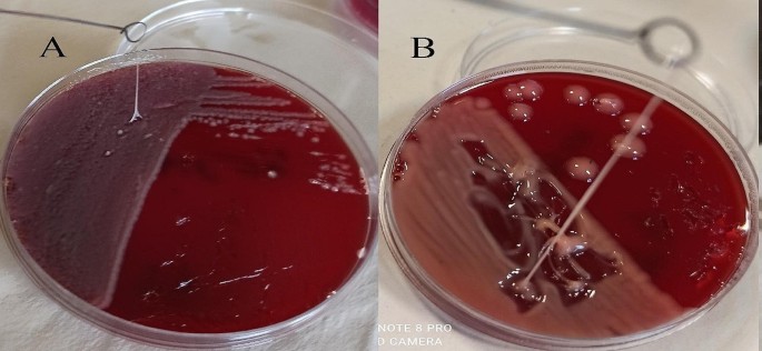
After two weeks, the patient had progressive constitutional symptoms and fever. Therefore, he was again referred to Abu Ali Sina Hospital and admitted to the post-transplant ward. In the subdiaphragmatic part of the right lobe of the liver, there was a large liver abscess and pigtail catheter drainage was performed. Biochemical tests also revealed a WBC count of 15,700/µL, BUN level of 40 mg/dL, Cr of 1.8 mg/dL, ALP of 1203 U/L, AST of 84 U/L, ALT of 119 U/L, D. bilirubin of 3.1 mg/dL, T. bilirubin of 5.39 mg/dL, and Hb of 13 g/dL (Table 1). Antibiotic treatment was started by administrating colistin 4.5 × 10⁶ units IV every 12 h and amikacin 500 mg IV every 12 h. Meanwhile, we isolated and identified two species of K. pneumoniae and E. coli in the pus culture. Here, the phenotypic and genotypic characteristics of the K. pneumoniae isolate had changed. On blood agar, the colonies were transparent with a sticky consistency and adhered to the culture medium (sticky mucoviscous colonies) (Fig. 1.A). The isolate was resistant to all tested antibiotics and was identified as a multidrug-resistant (MDR) isolate [15]. The minimum inhibitory concentration (MIC) of imipenem for K. pneumoniae was determined to be 128 µg/mL, by broth microdilution method according to CLSI recommendations [14]. MIC test for colistin was also performed by broth microdilution method according to CLSI guidelines [14] and the isolate was sensitive to colistin (MIC < 2 µg/mL). In addition, production of extended-spectrum β-lactamase (ESBL) and carbapenemase was detected by the phenotypic confirmatory disc diffusion test (PCDDT) and the modified carbapenem inactivation method (mCIM) according to CLSI guidelines [14]. Meanwhile, the E. coli isolate was also resistant to all tested antibiotics and was identified as an MDR isolate [15]. In K. pneumoniae and E. coli isolates, virulence genes entB, ybtS and antibiotic resistance genes blaTEM, blaSHV, blaCTX−M, and blaOXA−48 were detected by PCR using the specific primers [3] (Table 2). K. pneumoniae isolates belonged to sequence type 54 (ST54) by MLST and were identified as a non-K1/K2 ST54 cKp strain. On the 11th and 18th days of hospitalization, K. pneumoniae was again isolated and identified along with E. coli in pus culture. On the 20th day, despite the poor condition of the patient, he was discharged with personal consent and insistence and stated that he will continue his hospitalization and treatment in one of the hospitals in Tehran. The patient was advised that it is necessary to continue treatment with colistin 4.5 × 10⁶ units IV every 12 h and amikacin 500 mg IV every 12 h and to follow up the pigtail inserted into the liver abscess.
After 6 weeks, the patient was again referred with pneumocath and shingles and was admitted to the emergency department. The patient stated that he was hospitalized for SARS-CoV-2 infection two weeks ago. The primary diagnosis was pleural effusion, shingles, and liver abscess. On the first day, a CT scan of the chest revealed that all parts of both lung fields were clear without active pulmonary infiltrates or fibrocystic disease. The patient was alert, with stable vital signs, apyretic, vesicular skin lesions in the background of painful erythema (shingles) on the left side of the face and neck, clear lungs, and purulent discharge from the catheter exit site with a positive K. pneumoniae culture. The patient’s treatment continued with the administration of colistin, meropenem 500 mg IV every 12 h, and acyclovir. On the sixth day of hospitalization, the patient was alert, apyretic, ulcerative lesions on the face and neck were healing, clear lungs, and a pyogenic liver abscess of 200 × 130 × 61 mm with a positive culture of K. pneumoniae. Colistin, imipenem, doxycycline 100 mg every 12 h, and acyclovir were prescribed. Laboratory data revealed a WBC count of 10,200/µL, BUN level of 24 mg/dL, Cr of 1.5 mg/dL, ALP of 1416 U/L, AST of 30 U/L, ALT of 36 U/L, D. bilirubin of 0.6 mg/dL, T. bilirubin of 1 mg/dL, Hb of 7.4 g/dL (Table 1), and subsequently blood transfusion was prescribed to increase blood Hb. On the ninth day, while the liver abscess was very large, surgical drainage was performed. On the 11th day, the patient was alert, with stable vital signs, apyretic, and clear lungs. Purulent secretions were less and K. pneumoniae was detected in culture. The patient was receiving colistin, imipenem, tigecycline 50 mg IV every 12 h, fluconazole (used to treat fungal infections) 100 mg every 12 h, and acyclovir. Laboratory data revealed a WBC count of 6400/µL, BUN level of 32 mg/dL, Cr of 1.1 mg/dL, ALP of 1692 U/L, AST of 14 U/L, ALT of 19 U/L, D. bilirubin of 0.7 mg/dL, T. bilirubin of 1.4 mg/dL, Hb of 9.5 g/dL. On the 18th day, the patient was alert, with stable vital signs, apyretic, clear lungs, and shingles lesions were improving. The secretions were brief and K. pneumoniae was detected in the culture. Laboratory data revealed a WBC count of 6100/µL, BUN level of 14 mg/dL, Cr of 1.5 mg/dL, ALP of 1628 U/L, AST of 25 U/L, ALT of 45 U/L, D. bilirubin of 0.5 mg/dL, T. bilirubin of 0.9 mg/dL, Hb of 7.3 g/dL, and subsequently blood transfusion was prescribed to increase blood Hb. The patient was receiving colistin, doxycycline, and amikacin. On the 30th day, the patient was alert, with stable vital signs, apyretic, and K. pneumoniae was detected in the culture of the abscess secretions and Enterococcus sp. in the culture of the wound. On the 42nd day, the patient was alert, with stable vital signs, apyretic, and the shingles lesions were dry and improving. K. pneumoniae was detected in culture of liver abscess secretions. The patient was receiving colistin, tigecycline, doxycycline, fluconazole, and linezolid 600 mg every 12 h. On the 47th day, laboratory data revealed a WBC count of 7300/µL, BUN level of 15 mg/dL, Cr of 0.9 mg/dL, CRP of 8 mg/L, ALP of 4353 U/L, AST of 101 U/L, ALT of 208 U/L, D. bilirubin of 6 mg/dL, T. bilirubin of 12 mg/dL, and Hb of 7 g/dL (Table 1). The patient was discharged by personal desire, while all the professors were of the opinion that the patient should remain hospitalized. On the 60th day, the patient died while the clinical findings indicated that PLA was complicated by sepsis.
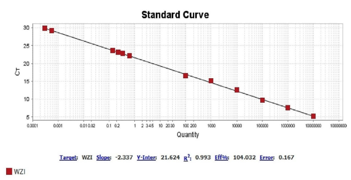
In order to investigate capsular polysaccharide expression, biofilm formation, and the presence of efflux pumps before and after imipenem treatment, a K1 ST23 hypervirulent K. pneumoniae (hvKp) strain, previously associated with invasive cryptogenic PLA [3], was included in the present study. HvKp produces a hypercapsule known as the hypermucoviscous phenotype, which is detected by a positive string test [3] (Fig. 1.B). The ability to form biofilm in two isolates sensitive (mucoviscous colonies) and CRKP (sticky mucoviscous colonies) isolated from non-cryptogenic PLA and hvKp isolate (hypermucoviscous colonies) isolated from cryptogenic PLA was investigated according to the previously described microtiter plate method [16]. All three K. pneumoniae isolates were biofilm producers. HvKp and sensitive cKp were identified as strong biofilm producers (optical density (OD) was 3.465 and 0.77, respectively), whereas CRKP was identified as a moderate biofilm producer (OD was 0.181) (Table 2). Interestingly, the biofilm formation ability of the hvKp strain was 16-fold higher than that of the positive control strain and almost 5-fold higher than that of sensitive cKp isolate, indicating a higher biofilm formation ability of hvKp compared to cKp. The presence of the efflux pump as an antibiotic resistance mechanism was also investigated according to the previously described ethidium bromide-agar cartwheel method [17]. The efflux pump mechanism could not be detected in all three isolates. Quantitative real-time PCR revealed that the expression of capsule wzi gene was nearly the same in hypermucoviscous and sticky mucoviscous isolates (cycle threshold (CT) was 22.47 and 23.12, respectively) and was approximately 1.3-fold that of the mucoviscous isolate (CT was 29.87) (Fig. 2).
A. Sticky mucoviscous colonies of CRKP isolate. B. Hypermucoviscous colonies and positive string test of ST23 hvKp strain. A positive string test is defined as the formation of a viscous string > 5 mm in length when colonies grown overnight on a blood agar plate at 37 °C are stretched with a bacteriological loop
Discussion and conclusions
Classical K. pneumoniae (cKp) is associated with non-cryptogenic PLAs in patients with underlying diseases including liver transplantation and diabetes mellitus [2, 3]. Global prevalence of CRKP is increasing, and colistin is considered the last resort antimicrobial to combat CRKP [8]. However, antibiotic resistance is a complex process and there is evidence that antibiotics promote antibiotic resistance [18]. Antibiotics damage bacterial cell structures and induce stress responses such as SOS and Rcs responses [10, 18]. Induction of stress responses leads to emergence of antibiotic resistance [18]. At this time, the role of administration of carbapenems in promoting colistin resistance in K. pneumoniae is unknown.
In this study, we report the evolution of a carbapenem-susceptible K. pneumoniae strain to CRKP by acquiring blaOXA−48 during imipenem treatment. Our clinical case highlights the possibility of horizontal gene transfer (HGT) between species during infection. HGT is the major mechanism responsible for the spread of carbapenemase genes through plasmids, transposons, and integrons among bacterial species [19, 20]. As previously reported, several classes of antibiotics (e.g. fluoroquinolones, β-lactams, and aminoglycosides) induce the SOS response at sub-MIC levels. The downstream consequences of induction can lead to the emergence of antibiotic resistance by mutations, biofilm formation, and the development of HGT [18, 21]. This study also showed that imipenem may be a promoter of resistance to carbapenems in K. pneumoniae. Unfortunately, due to the limitations of the present study, whole genome sequencing was not performed.
K. pneumoniae can acquire resistance to colistin by modifying LPS through addition of cationic groups to lipid A. LPS modification is mainly acquired through chromosomal mutations in the crrA/crrB or mgrB genes [22]. In addition, K. pneumoniae increases the amount of capsular polysaccharide and upregulates the transcription of the cps operon when grown in vitro in the presence of polymyxin B, leading to increased polymyxin resistance [23]. In vitro studies have shown that exposure to sub-MIC concentrations of β-lactams such as imipenem, which target peptidoglycan, can increase biofilm formation, whereas antibiotics that target the ribosome or DNA replication are less likely to do so [24, 25]. K. pneumoniae typically infects patients with indwelling medical devices such as catheters, on which the bacterium can grow as a biofilm [26]. Biofilm development begins with the reversible attachment of planktonic bacteria to a surface, and in the next step, the attachment becomes irreversible and the bacteria multiply and form microcolonies on the surface that begin to produce extracellular polysaccharides (EPS) around the microcolonies [12, 27]. EPS are an insoluble and slimy secretion that are released by bacterial cells and lead to biofilm formation [12]. Bacterial biofilms are a serious problem in medicine because they cause chronic infections due to their high resistance to antibiotics and the host’s defense system [27]. The biofilm structure prevents the penetration of antibiotics. Furthermore, within biofilms, persister cells that are temporarily dormant or grow very slowly are associated with the emergence of antibiotic resistance [27]. According to published research, resistant bacterial cells increase the minimum inhibitory concentration (MIC), whereas no increase in MIC is observed in persister cells [28, 29]. Meanwhile, the ability of invasive hvKp strains to form biofilms is significantly higher than that of non-invasive cKp isolates [30]. In hvKp strains, rmpA (regulator of mucoid phenotype) and rmpA2 genes increase the expression of cps genes, resulting in a hypermucoviscous phenotype [31, 32]. An increase in the amount of capsular polysaccharide contributes to the formation of a stronger biofilm in K. pneumoniae [30, 33]. But why was the biofilm-forming ability of the CRKP (sticky mucoviscous colonies) isolate, which overexpressed the cps operon almost as much as the hvKp strain, reduced to a moderate level? Interestingly, no hypermucoviscous phenotype was also observed in this isolate. Extracellular polysaccharides (EPS) are known as capsule, slime, and glycocalyx [34]. The distinction between capsule and slime is not clear, and glycocalyx is usually used as a general term to refer to EPS [34]. The capsule is tightly bound to the bacterial cell and is not removable by repeated washing, whereas the slime layer is loosely bound to the bacterial cell and is easily washed off [34, 35]. Slime also imparts a sticky consistency to bacterial growth on a solid medium [34, 36]. We observed that CRKP colonies on blood agar had a sticky consistency and adhered to the culture medium. In addition, the capsule was loose and easily washed off during washing, which likely reduced biofilm formation in the biofilm formation assay.
Slime-producing bacterial strains grow embedded in an insoluble glucan matrix associated with surfaces and form very thick biofilms compared to non-slime-producing strains that preferentially grow as non-adherent cells in the culture supernatant [35]. Therefore, slime is involved not only in increasing the initial adhesion, but also in the subsequent formation of microcolonies [35]. The present study shows that imipenem may increase capsular polysaccharide expression and slime production in CRKP strains, which can exacerbate biofilm formation in PLA catheters and promote colistin resistance. It would be of concern if antimicrobial agents used to treat infections exacerbated biofilm formation, thereby protecting bacteria from antimicrobial agents.
Colistin is widely used to treat intra-abdominal infections and sepsis by CRKP [5]. However, the implementation of colistin monotherapy against these infections is associated with the negative outcome of the emergence of colistin-resistant CRKP [5]. Therefore, colistin is usually prescribed in combination therapy protocols with fosfomycin, tigecycline, carbapenems, and aminoglycosides. Combinations of colistin increase their bactericidal effect against CRKP isolates [5, 37]. In addition, cefiderocol, imipenem cilastatin/relebactam, meropenem-vaborbactam, ceftazidime–avibactam, and aztreonam–avibactam are potent alternatives for the treatment of CRKP infections [5]. At the time of the present study, none of these new antibiotics were available in our country.
The limitations of the present study were that the physician’s choice to prescribe certain antibiotics was often unclear, the doubt that the patient followed the treatment in the period after voluntary discharge, and finally, the whole bacterial genome could not be sequenced.
In this case, which was an immunocompromised patient (diabetes mellitus and immunosuppressive drugs), the K. pneumoniae infection was not controlled and the patient died. Factors that can be involved in the emergence of antibiotic resistance and the death of this patient: (A) Treatment with inadequate dose of imipenem and emergence of resistance to carbapenems. (B) Low level of immunity and possibility of opportunistic infections. (C) Sudden changes in prescribed antibiotics. (D) The possibility of mixed infection from the beginning. (E) The patient’s involvement in the treatment process due to the problems caused by being away from the place of residence..
Based on this information, we have a theoretical hypothesis that imipenem is a promoter of resistance to carbapenems and colistin in K. pneumoniae. This needs more attention.
Data availability
All relevant data are fully presented in the manuscript.
Abbreviations
- PLA:
-
Pyogenic liver abscess
- MDR:
-
Multidrug-resistant
- CRKP:
-
Carbapenem-resistant K. pneumoniae
- EPS:
-
Extracellular polysaccharides
- ESBL:
-
Extended-spectrum-β-lactamase
- PCDDT:
-
Phenotypic confirmatory disk diffusion test
- Mcim:
-
Modified carbapenem inactivation method
- ST:
-
Sequence type
- HGT:
-
Horizontal gene transfer
References
Lardière-Deguelte S, Ragot E, Amroun K, Piardi T, Dokmak S, Bruno O, et al. Hepatic abscess: diagnosis and management. J Visc Surg. 2015;152(4):231–43.
Paczosa MK, Mecsas J. Klebsiella pneumoniae: going on the offense with a strong defense. Microbiol Mol Biol Rev. 2016;80(3):629–61.
Sohrabi M, Alizade Naini M, Rasekhi A, Oloomi M, Moradhaseli F, Ayoub A, et al. Emergence of K1 ST23 and K2 ST65 hypervirulent Klebsiella pneumoniae as true pathogens with specific virulence genes in cryptogenic pyogenic liver abscesses Shiraz Iran. Front cell Infect. 2022;12:964290.
Mavilia MG, Molina M, Wu GY. The evolving nature of hepatic abscess: a review. J clin Transl Hepatol. 2016;4(2):158.
Karampatakis T, Tsergouli K, Behzadi P. Carbapenem-resistant Klebsiella pneumoniae: virulence factors, molecular epidemiology and latest updates in treatment options. Antibiotics. 2023;12(2):234.
Lee C-R, Lee JH, Park KS, Kim YB, Jeong BC, Lee SH. Global dissemination of carbapenemase-producing Klebsiella pneumoniae: epidemiology, genetic context, treatment options, and detection methods. Front Microbiol. 2016:895.
Pitout JD, Peirano G, Kock MM, Strydom K-A, Matsumura Y. The global ascendency of OXA-48-type carbapenemases. Clin Microbiol Rev. 2019;33(1). https://doi.org/10.1128/cmr. 00102– 19.
Petrosillo N, Taglietti F, Granata G. Treatment options for colistin resistant Klebsiella pneumoniae: present and future. J Clin Med. 2019;8(7):934.
Zhu J, Wang T, Chen L, Du H. Virulence factors in hypervirulent Klebsiella pneumoniae. Front Microbiol. 2021;12:642484.
Meng J, Young G, Chen J. The Rcs system in Enterobacteriaceae: envelope stress responses and virulence regulation. Front Microbiol. 2021;12:627104.
Sailer FC, Meberg BM, Young KD. β-Lactam induction of colanic acid gene expression in Escherichia coli. FEMS Microbiol Lett. 2003;226(2):245–9.
Veerachamy S, Yarlagadda T, Manivasagam G, Yarlagadda PK. Bacterial adherence and biofilm formation on medical implants: a review. Proc Inst Mech Eng H: J Eng Med. 2014;228(10):1083–99.
Mahon CR, Lehman DC. Textbook of diagnostic microbiology-e-book. Elsevier Health Sciences; 2022.
Weinstein M, Patel J, Bobenchik A, Campeau S, Cullen S, Galas M et al. M100 Performance standards for Antimicrobial susceptibility testing a CLSI supplement for global application. Performance standards for antimicrobial susceptibility testing performance standards for antimicrobial susceptibility testing. 2020.
Magiorakos A-P, Srinivasan A, Carey RB, Carmeli Y, Falagas M, Giske C, et al. Multidrug-resistant, extensively drug-resistant and pandrug-resistant bacteria: an international expert proposal for interim standard definitions for acquired resistance. Clin Microbiol Infect. 2012;18(3):268–81.
Nirwati H, Sinanjung K, Fahrunissa F, Wijaya F, Napitupulu S, Hati VP, et al. editors. Biofilm formation and antibiotic resistance of Klebsiella pneumoniae isolated from clinical samples in a tertiary care hospital, Klaten, Indonesia. BMC Proc; 2019.
Kareem SM, Al-Kadmy IM, Kazaal SS, Ali ANM, Aziz SN, Makharita RR, et al. Detection of gyra and parc mutations and prevalence of plasmid-mediated quinolone resistance genes in Klebsiella pneumoniae. Infect Drug Resist. 2021;14:555.
Laureti L, Matic I, Gutierrez A. Bacterial responses and genome instability induced by subinhibitory concentrations of antibiotics. Antibiotics. 2013;2(1):100–14.
Ding B, Shen Z, Hu F, Ye M, Xu X, Guo Q, et al. In vivo acquisition of carbapenemase gene bla KPC-2 in multiple species of Enterobacteriaceae through horizontal transfer of insertion sequence or plasmid. Front Microbiol. 2016;7:1651.
Hamprecht A, Sommer J, Willmann M, Brender C, Stelzer Y, Krause FF, et al. Pathogenicity of clinical OXA-48 isolates and impact of the OXA-48 IncL plasmid on virulence and bacterial fitness. Front Microbiol. 2019;10:2509.
Andersson DI, Hughes D. Microbiological effects of sublethal levels of antibiotics. Nat Rev Microbiol. 2014;12(7):465–78.
Wang Y, Luo Q, Xiao T, Zhu Y, Xiao Y. Impact of polymyxin resistance on virulence and fitness among clinically important gram-negative bacteria. Engineering. 2022;13:178–85.
Olaitan AO, Morand S, Rolain J-M. Mechanisms of polymyxin resistance: acquired and intrinsic resistance in bacteria. Front Microbiol. 2014;5:643.
Ranieri MR, Whitchurch CB, Burrows LL. Mechanisms of biofilm stimulation by subinhibitory concentrations of antimicrobials. Curr Opin Microbiol. 2018;45:164–9.
Nucleo E, Steffanoni L, Fugazza G, Migliavacca R, Giacobone E, Navarra A, et al. Growth in glucose-based medium and exposure to subinhibitory concentrations of imipenem induce biofilm formation in a multidrug-resistant clinical isolate of Acinetobacter baumannii. BMC Microbiol. 2009;9:1–14.
Singla S, Harjai K, Chhibber S. Susceptibility of different phases of biofilm of Klebsiella pneumoniae to three different antibiotics. J Antibiot. 2013;66(2):61–6.
Høiby N, Bjarnsholt T, Givskov M, Molin S, Ciofu O. Antibiotic resistance of bacterial biofilms. Int J Antimicrob Agents. 2010;35(4):322–32.
Algammal A, Hetta HF, Mabrok M, Behzadi P. Emerging multidrug-resistant bacterial pathogens superbugs: a rising public health threat. Front Microbiol. 2023;14:1135614.
Brauner A, Fridman O, Gefen O, Balaban NQ. Distinguishing between resistance, tolerance and persistence to antibiotic treatment. Nat Rev Microbiol. 2016;14(5):320–30.
Wu M-C, Lin T-L, Hsieh P-F, Yang H-C, Wang J-T. Isolation of genes involved in biofilm formation of a Klebsiella pneumoniae strain causing pyogenic liver abscess. PLoS ONE. 2011;6(8):e23500.
Lai Y-C, Peng H-L, Chang H-Y. RmpA2, an activator of capsule biosynthesis in Klebsiella pneumoniae CG43, regulates K2 cps gene expression at the transcriptional level. J Bacteriol. 2003;185(3):788–800.
Lee C-R, Lee JH, Park KS, Jeon JH, Kim YB, Cha C-J, et al. Antimicrobial resistance of hypervirulent Klebsiella pneumoniae: epidemiology, hypervirulence-associated determinants, and resistance mechanisms. Front Cell Infect Microbiol. 2017;7:483.
Dzul SP, Thornton MM, Hohne DN, Stewart EJ, Shah AA, Bortz DM, et al. Contribution of the Klebsiella pneumoniae capsule to bacterial aggregate and biofilm microstructures. Appl Environ Microbiol. 2011;77(5):1777–82.
Drewry DT, Galbraith L, Wilkinson BJ, Wilkinson SG. Staphylococcal slime: a cautionary tale. J Clin Microbiol. 1990;28(6):1292–6.
Baselga R, Albizu I, Amorena B. Staphylococcus aureus capsule and slime as virulence factors in ruminant mastitis. A review. Vet Microbiol. 1994;39(3–4):195–204.
Wilkinson J. The extracellular polysaccharides of bacteria. Bacteriol rev. 1958;22(1):46–73.
Yu L, Zhang J, Fu Y, Zhao Y, Wang Y, Zhao J, et al. Synergetic effects of combined treatment of colistin with meropenem or amikacin on carbapenem-resistant Klebsiella pneumoniae in vitro. Front Cell Infect Microbiol. 2019;9:422.
Acknowledgements
The authors would like to thank the staff of the Department of Bacteriology and Virology of Shiraz University of Medical Sciences, Abu Ali Sina Hospital, Fara Parto Medical Imaging and Radiology Center, and Pasteur Institute of Iran. This research was supported by the Pasteur Institute of Iran (Project No.: B-9636).
Funding
Funding was granted to Maryam Sohrabi as a Ph.D. student of the Pasteur Institute of Iran (Project No.: B-9636).
Author information
Authors and Affiliations
Contributions
MS & NP performed the phenotypic and genotypic tests. MS collected the clinical data. MS wrote the manuscript. NP & MA wrote and revised the manuscript. AR & AA collected the samples. MS and ZH analyzed the data. FS supervised the project and wrote and revised the manuscript. All authors read and approved the final manuscript.
Corresponding author
Ethics declarations
Ethics approval and consent to participate
The studies involving human participants were reviewed and approved by the ethics committee of the Pasteur Institute of Iran (reference number: IR.PII. REC.1399.065). Written informed consent to participate in this study was provided by the patient.
Consent for publication
Written informed consent was obtained from the patient to publish this case report.
Competing interests
The authors declare that they have no competing interests.
Additional information
Publisher’s Note
Springer Nature remains neutral with regard to jurisdictional claims in published maps and institutional affiliations.
Rights and permissions
Open Access This article is licensed under a Creative Commons Attribution 4.0 International License, which permits use, sharing, adaptation, distribution and reproduction in any medium or format, as long as you give appropriate credit to the original author(s) and the source, provide a link to the Creative Commons licence, and indicate if changes were made. The images or other third party material in this article are included in the article’s Creative Commons licence, unless indicated otherwise in a credit line to the material. If material is not included in the article’s Creative Commons licence and your intended use is not permitted by statutory regulation or exceeds the permitted use, you will need to obtain permission directly from the copyright holder. To view a copy of this licence, visit http://creativecommons.org/licenses/by/4.0/. The Creative Commons Public Domain Dedication waiver (http://creativecommons.org/publicdomain/zero/1.0/) applies to the data made available in this article, unless otherwise stated in a credit line to the data.
About this article
Cite this article
Sohrabi, M., Pirbonyeh, N., Alizade Naini, M. et al. A challenging case of carbapenem resistant Klebsiella pneumoniae-related pyogenic liver abscess with capsular polysaccharide hyperproduction: a case report. BMC Infect Dis 24, 433 (2024). https://doi.org/10.1186/s12879-024-09314-z
Received:
Accepted:
Published:
DOI: https://doi.org/10.1186/s12879-024-09314-z

